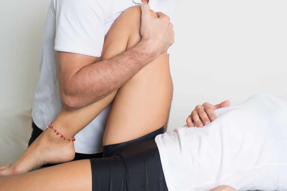
Part 2 of our ACL blog series will include a review of screening tools and preventative interventions for patients that may be "at risk" for an ACL tear. If you have not checked out 'Part 1: Structure and Function', you can find it here: Anterior Cruciate Ligament Part I or on our Facebook page!
As we learned in Part 1 of this series, around 70% of all ACL ruptures occur due to non-contact injuries, or those sustained due to tissue failure. These tears can be due to a structural deficit (angle of alignment between the femur and tibia) or a muscular strength deficit that is caused by too much force to be transmitted through the ACL and resulting in rupture. Below are screening tools that can be used by physical therapists and other qualified movement professionals to assess whether an athlete has structural deficiencies or movement patterns that would predispose them to an ACL rupture.
- Q-angle: This is the angle created between a line from the Anterior Superior Iliac Spine (ASIS) and the midpoint of the patella (kneecap) and a line from the midpoint of the kneecap (patella) through the tibial tuberosity (large bump on the front of the shin bone). This angle is usually greater in females, as they are more likely to have wider hips and a valgus knee (knees sit closer to midline than the ankle). The average Q-angle is 13.5 degrees for men and 18.1 degrees for women.
- Single-Leg Step Down: The single-leg step down involves lowering from a step using only one leg, tapping the opposite foot on the ground, and returning to the starting position. This motion allows the practitioner to take note of how the athlete or client uses various muscles to complete a movement pattern. By having the client perform a step down (opposed to a step up), more insight is given to eccentric control of the muscles. An eccentric contraction occurs when the muscle is producing force that lengthens rather than shortens.
- Weight-bearing Ankle Mobility: Ankle mobility is one of three movements that have been linked to risk of ACL rupture. When measured in weight-bearing, there is more insight to what is happening at the joint when the foot is planted, as it is in the stance phase of running. This can be measured by having the foot planted and heel remaining in contact with the floor, then bending the knee and sliding it forward until a restriction is felt in the calfs.
- Overhead Deep Squat: Using the overhead deep squat as a screening tool allows the clinician to evaluate upper and lower extremity movement patterns simultaneously. This movement requires lower extremity flexion with upper extremity extension, requiring significant core muscle control because the muscles of the trunk and proximal joints (hips and shoulders) must stabilize, Common compensations that occur during this movement include anterior collapse at the upper trunk, posterior collapse of the shoulder joint, collapse at the pelvis, use of one extremity more than the other while lowering or returning to stand, inability to sink down completely due to a tight posterior chain or inability to push the head of the femur to the back of the hip socket. Each of these compensations can present in different ways, which would involve too extensive a list to include here but awareness of these issues can help.
- Tuck Jump: The tuck jump is a more dynamic movement that requires both take-off and landing by the client. Among other qualities, the clinician will be able to determine muscle versus ligament dominance in stabilization during the countermovement (squat before the jump), the ability to load hips eccentrically to develop and store power, quad or glute dominance with take-off, core control when in the air and up landing, as well as hip stability, loading ability, ankle mobility, and muscle balance during landing.
Although this is not a complete list of the screening tools available when looking at the risks of ACL tears, these tests give an idea of the movements and patterns associated with an injury. Following the screening, the clinician will be able to have a picture of the deficits present, and be able to tailor a program to address the problem list. Periodically throughout the program, subsequent screenings should take place to measure efficiency and effectiveness of the implemented program. It is also important that the tests or screens themselves are not part of the exercise program, as it has been supported in the literature that clients are able to improve those scores while maintaining a deficit.
If you think you may be at risk for an ACL tear, you can always reach out to your SetPT physical therapist for more information on a screening. Stay tuned for Part 3: ACL Reconstruction Rehabilitation coming soon!
Article Abstracts:
Single-Leg Step Down: http://www.jospt.org/doi/abs/10.2519/jospt.2007.2202#.VjkLGNKrTDc
Ankle Dorsiflexion and Landing Biomechanics: http://www.ncbi.nlm.nih.gov/pubmed/21214345
Weight-bearing versus nonweight-bearing ankle dorsiflexion measurements: http://search.proquest.com/openview/e678ac1437651495f9894f11307c1d30/1?pq-origsite=gscholar
Tuck Jumps and Effectiveness when Used in Prevention Screening: http://journals.lww.com/nsca-jscr/Abstract/2011/03001/The_Effects_of_Pre_Season_and_In_Season.192.aspx


